
Tel: (+86)15251899190
WeChat: KJ15251899190
E-mail: 2853036907@qq.com
Address: Complex building Lev11,
Jingang Sci-Tech Center, Nanjing.

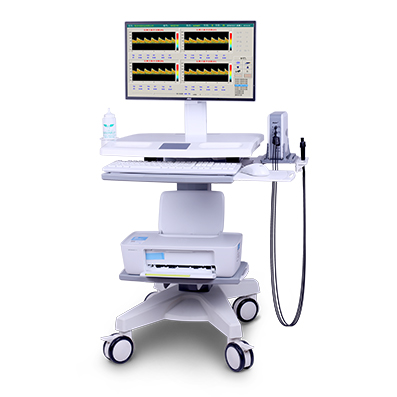
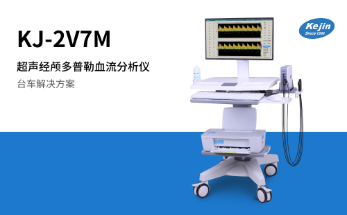
Product Name: Ultrasound Transcranial Doppler Blood Flow Analyzer
Product model: KJ-2V7M
Detection principle: Ultrasonic Doppler principle
Domestic/Imported: Domestic
Medical/Household: Medical
Available for sale: nationwide
Input power: 60VA
Operating temperature: 5-40 ℃
Operating humidity: Relative humidity ≤ 80%
The following product introductions are all about the overall solution of the product


The KJ-2V7M ultrasound transcranial Doppler blood flow analyzer integrates multiple TCD technologies such as small signal recognition, M-mode, and automatic vascular search. It can achieve single and dual channel dual-mode detection and is equipped with a composite material ultrasound probe. The multi depth mode can simultaneously display blood flow spectrum signals at eight depths.
The KJ-2V7M TCD instrument is equipped with two KJ-PW-2MHz probes, one KJ-CW-4MHz probe, and one KJ-CW-8MHz probe, which can be used for intracranial vascular detection, extracranial vascular detection, and limb capillary detection. The overall solution of this product is equipped with a LCD computer all-in-one machine, a convenient trolley, and a color inkjet printer, which can conveniently achieve the detection function of an ultrasound transcranial Doppler blood flow analyzer.
Adaptation department
Neurology, neurosurgery, intensive care unit, functional examination department, physical examination department, etc.
Functional characteristics
1. Ultrasonic probe technology
By using composite ceramic material chips, matching layer technology, and cutting processes, the signal-to-noise ratio and energy efficiency ratio of the sound beam have been improved, ensuring ultrasound penetration and resolution of ultrasound images. Imitation of German probe structure, with high signal-to-noise ratio and sensitivity.
Multi probe configuration, including KJ-CW-8MHz probe, can be used for detecting carotid arteries and small blood vessels in limbs, expanding the application field of the product, facilitating various examinations, and improving the investment value of the equipment.
2. Weak signal enhancement
Wavelet processing technology enhances weak signals, uses intelligent algorithms to analyze and process ultrasound echo signals, improves detection sensitivity, spectrum image display clarity, and makes it easier to find vascular signals.
3. M module function
The M-mode function can simultaneously display multiple blood flow signals of different depths and quickly locate the depth of blood flow, helping doctors obtain blood flow spectrograms.
4. HITS detection technology
Embolism detection technology, when a embolus (HITS) appears, the signal displays a jumping waveform and can be replayed for recognition, making it easy for doctors to observe and detect.
5. Automatic vascular search function
The automatic blood vessel search technology captures weak signals, quickly locates the depth of blood vessels, shortens the time to search for the location of blood vessels, and reduces the dependence on the operator's high technical skills.
6. Multi channel and multi depth detection
Simultaneously displaying multiple depths of a blood vessel, identifying typical symptoms such as vascular stenosis, and observing the trajectory of thrombus flow; Dual channel spectrum display comparison provides a basis for evaluating bilateral vascular blood flow.
7. Information management and transmission
Quickly record patient information, the software includes workstation functions to manage all patient information, access patient detection data and reports at any time, and implement multimedia image management through DICOM 3.0 interface, wired and wireless network transmission, and USB interface.
8. Other technologies
1. Dynamic and static envelope lines, real-time display of spectrum, calculation of blood flow parameters;
2. Multiple reports are available for easy modification;
3. Check medical records at any time, store vascular blood flow spectra for movie playback;
9. Clinical application
1. Used for blood flow measurement of transcranial, cervical, and peripheral blood vessels, excluding analytical and diagnostic functions;
2. Detect the blood flow of blood vessels in the brain;
3. Detection of blood flow within superficial blood vessels;
4. Quantitative evaluation of the blood supply status of cerebral and superficial vessels can provide a basis for screening the nature, degree, and scope of various cerebrovascular diseases.
Advice: Please carefully read the product manual or purchase and use it under the guidance of medical personnel. Please refer to the manual for any contraindications or precautions.